4. Green Pigments: Exploring Changes in the Egyptian Color Palette through the Technical Study of Roman-Period Mummy Shrouds
This project began with a chance discovery: a saturated, soft blue-green background on a limestone funerary stela from the Roman Egyptian town of Terenouthis. Surprisingly, the color was not the same copper-based green found in a bowl also excavated at the site; rather, it was celadonite, the mineral (see fig. 4.1).1 Most publications on Egyptian materials and techniques provided little information on the use of green earth in Egypt, focusing primarily on materials used during the dynastic periods, when green were not employed. Only a small number of case studies cited the use of green earth on Egyptian artifacts. Curious to discover if there were other instances, I set out on a multiyear research project to study the use of green pigments in Greco-Roman Egyptian art. This work included an extensive review of existing literature on green pigments and a technical survey of artifacts in museum collections, including several at the J. Paul Getty Museum and the Metropolitan Museum of Art. The overarching goal of the research was to build a green pigment data set that was more inclusive of the Ptolemaic and Roman periods and to explore changes in pigment use in Egypt over time.
| Possible Pigments | Chemical Formulas | Description |
|---|---|---|
| Malachite Verdigris | CuCO3 Cu(OH)2 Cu(CH3COO)2 | Basic copper carbonate and copper acetate minerals |
| Chrysocolla | (Cu,Al)2H2Si2O5(OH)4·nH2O | A naturally occurring copper silicate mineral that often occurs with frit |
| Basic copper chlorides | Cu2Cl(OH)3 | Described as synthetic pigments in some publications and elsewhere as the alteration products of Egyptian blue and Egyptian green |
| Organo-copper compounds | Cu-proteinate, Cu-carbohydrate, and Cu-wax pigments | Possible reaction between altered copper-containing pigments and binding media |
| Egyptian blue Egyptian green | CaCuSi4O10 Cu glass | Synthetic blue and green pigments produced by heating sand, natron, flux, and copper minerals |
| Green earth | K(Mg,Fe2+)(Fe3+,Al)[Si2O10](OH)2 (K,Na)(Fe3+,Al,Mg)2(Si,Al)4O10 | A naturally occurring clay mineral—celadonite and glauconite are the two primary mineral species referenced |
| Mixtures | See description | Combinations of Egyptian blue (CaCuSi4O10), indigo, Egyptian green, orpiment (As2S3), yellow ochre (Fe2O3·H2O), and green earth |
Literature Survey
The literature review focused deliberately on the Ptolemaic and Roman periods—at least initially. To compare pigment use during these periods to that of earlier times, the project eventually expanded to include studies focused on dynastic Egypt. A variety of published sources were consulted as well, as were analytical results reported in the APPEAR database. Artifact names and , institutions and authors, and pigment characterizations were recorded. The review revealed that a wide range of materials had been used to create the color green in Egyptian art. Copper-based green pigments were reported most frequently and included minerals such as and verdigris,2 synthetic pigments such as Egyptian green,3 and alteration products such as copper chlorides and organo-copper compounds.4 Green earths and mixtures of blue and yellow pigments were also reported.5 A summary of these pigments is provided in figure 4.1.
Technical Survey
With the help of many collaborators, a technical survey of funerary artifacts was conducted within several Egyptian collections. Artifacts included inscribed stelae, coffins, cartonnage fragments, and painted mummy shrouds. The goal of the survey was to characterize green pigments on artifacts from Ptolemaic and Roman Egypt in particular and, in doing so, increase the number of existing case studies. The results of this survey reflected what was reported in the literature—with two important exceptions. First, green earth appeared more frequently than copper-based greens on Roman-period artifacts. Second, a blue-yellow pigment mixture, which has been cited in two previous publications,6 was found on at least two (possibly three) artifacts. These findings pointed to changes in pigment selection during the Ptolemaic and Roman periods—observations that will be discussed in the conclusions section below. Among the artifacts studied, it was the shrouds that yielded the most interesting results in terms of green pigments identified. The lack of copper greens, along with evidence of an expanded green palette, make the shrouds the most compelling case studies in terms of green color use.
Mummy Shrouds Case Study
The shrouds (figs. 4.2–4.8) are part of the collections of the J. Paul Getty Museum and the Metropolitan Museum of Art.7 Like portraits, painted textile mummy shrouds were a form of burial dress for the dead. The shrouds functioned both as part of the mummy’s encasement and as a surface for paint decoration. Sheets of textile were painted with portraits of the deceased; images, symbols, and writing from both Greek and Egyptian cultural traditions were also included. The portraits are at times naturalistic, depicting individuals as they were in life, but they can also be highly stylized, portraying the deceased as an embodiment of an Egyptian deity.8
Although the majority of shrouds in museum collections have been separated from the body, their overall shape, positioning of painted images and framing elements, wear patterns, and staining attest to their original use as mummy wrappings.9 The shrouds discussed in this paper, although fragmented, represent a number of portrait formats. These include the bust-length vignette format, in which the portrait is limited to the individual’s upper body; the full-length portrait; the three-figure format, in which the deceased is flanked by deities such as Osiris and Anubis; and shrouds in which the deceased is portrayed as a deity.10 The sections that follow will briefly describe each shroud.
Getty Museum
Two vignette-type shrouds from the J. Paul Getty Museum were analyzed. The painted portrait of a boy depicts a young man with a falcon on his left shoulder (see fig. 4.2). The painting ends just below the youth’s shoulders, indicating that the shroud would have been placed over the face of the deceased and secured with wrappings in the same manner as a portrait panel. The green of the leaves in the is strikingly vivid, and upon close examination one can observe bright yellow pigment particles, indicating that the paint is some kind of mixture.11
The painted portrait of a youth features a boy holding a floral and a cluster of grapes (see fig. 4.3). A falcon and a mummiform figure appear over his proper right shoulder. Visible in the wing to the boy’s left is a stripe of green; it appears to have been rendered by layering blue pigment over yellow pigment.
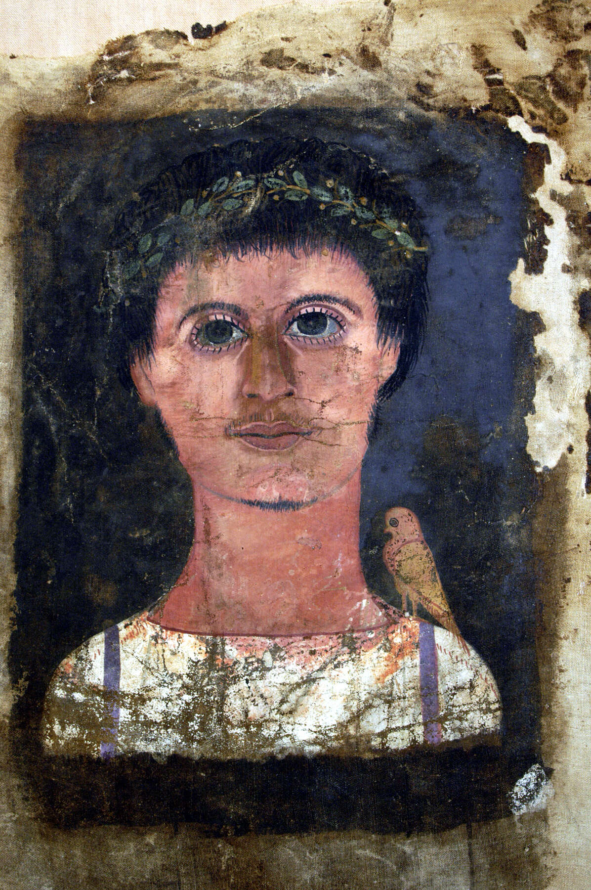 Figure 4.2
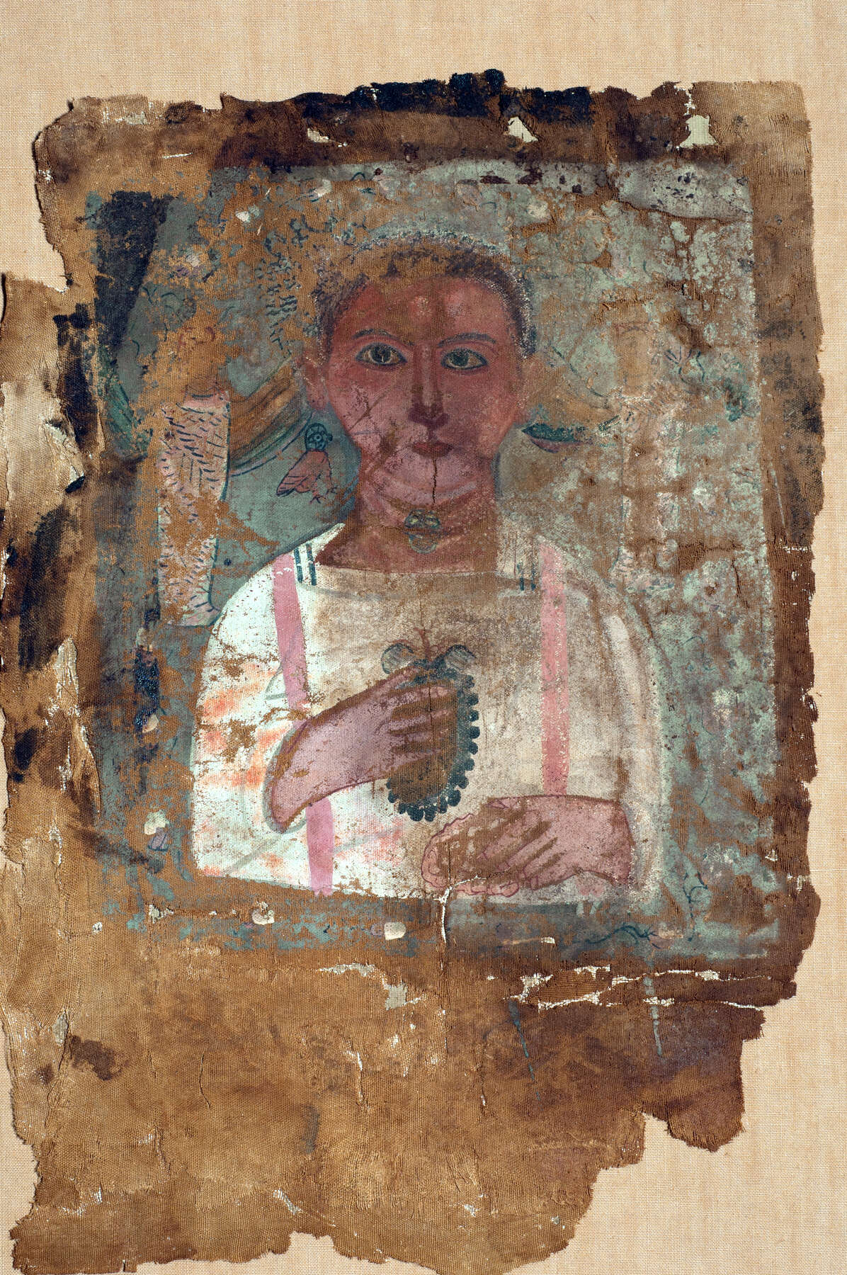 Figure 4.3
Metropolitan Museum
Four painted shroud fragments were analyzed at the Metropolitan Museum of Art. One example is a fragment of what was likely a full-length portrait shroud of a woman (see fig. 4.4). Only the woman’s beringed hands and a pink floral garland remain. The leaves in the garland are rendered in dark blue or black, pale blue, and green.
Another example is a full-length portrait shroud of a young man who takes the form of Osiris (see fig. 4.5). The figure is enshrined within an architectural framework crowned by an uraeus. Six adjacent registers depict Horus, Anubis, Ra-Horakhty, and a portrait of the deceased.12 The figure’s crown and crook as well as two of the hieroglyph registers are rendered in a pale turquoise color.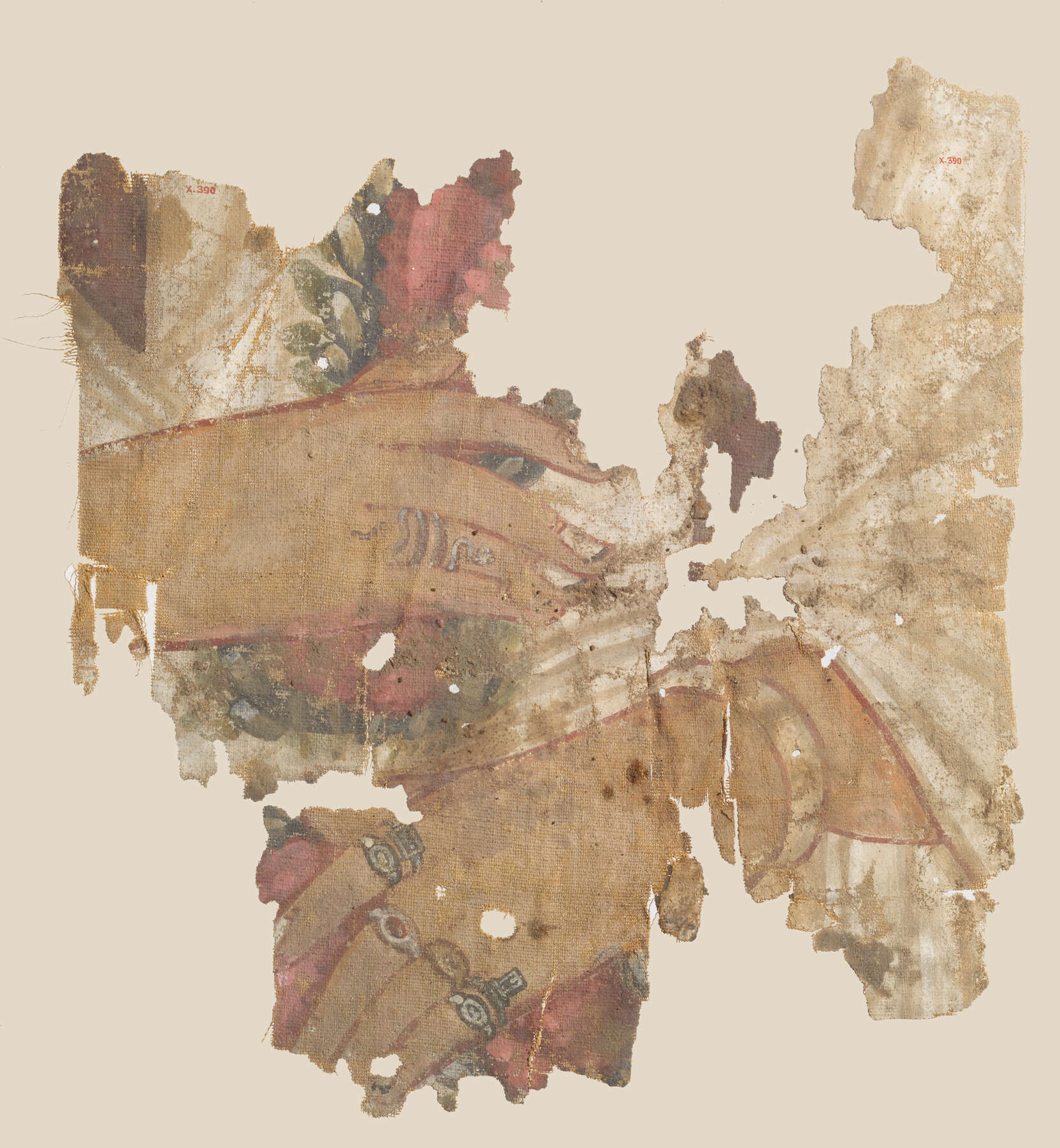 Figure 4.4
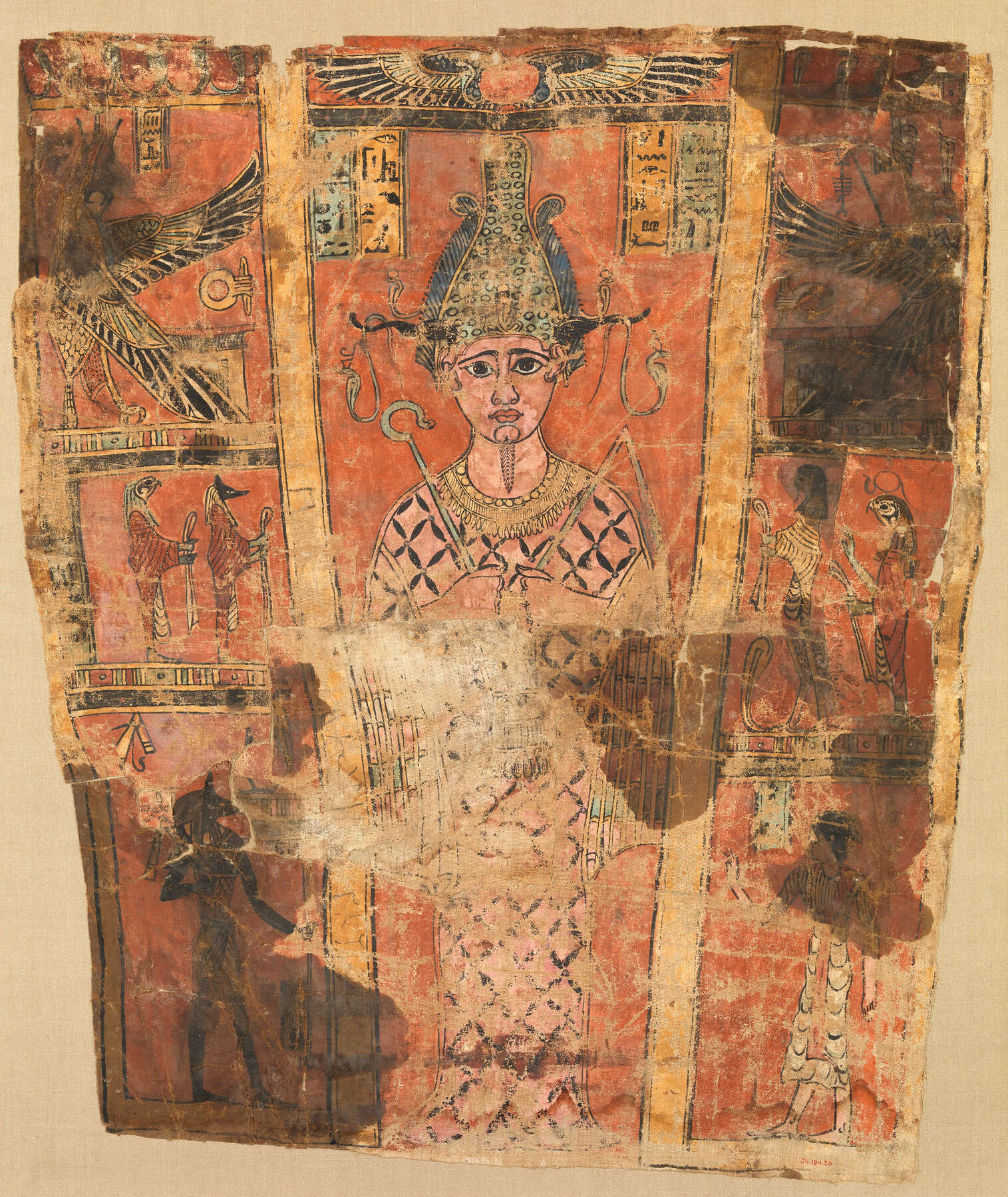 Figure 4.5
Two other fragments come from what appears to be an Osiris shroud. Fragment A (see fig. 4.6) shows the right shoulder of a mummiform figure covered with a bead net. The figure wears a broad collar and a wadjet eye amulet, and an unknown geometric form sits on the netting. Fragment B (see fig. 4.7) portrays a falcon head. The shroud fragments’ backgrounds are a colorful display of checkered patterning. One checkered section is rendered in yellow-green and dark green—the same dark green seen in the upper portion of the broad collar, in the unidentified geometric form, and below the falcon. The yellow-green checkerboard appears to have been made by superimposing yellow squares over a dark green background.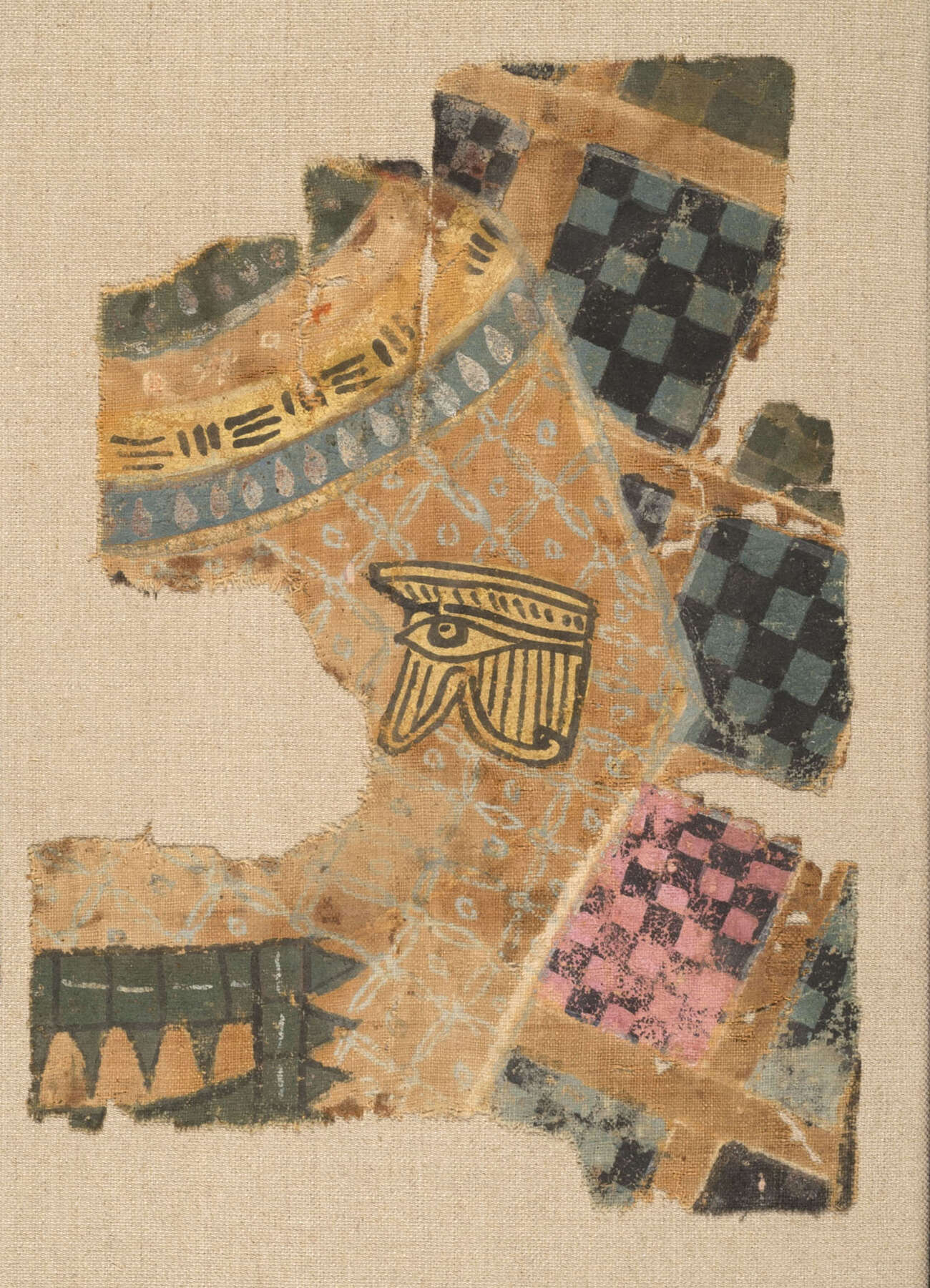 Figure 4.6
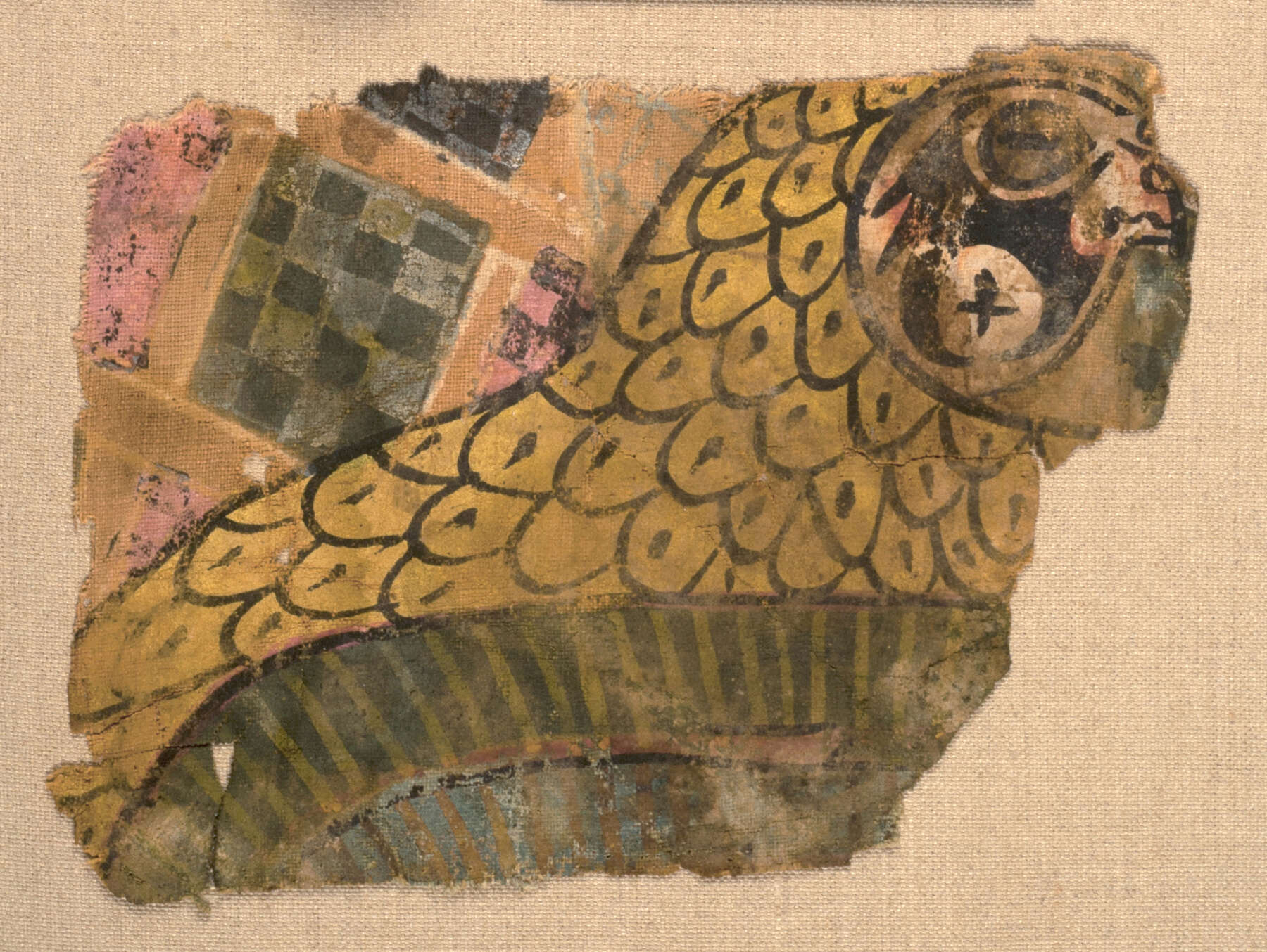 Figure 4.7
Two painted shroud fragments (66.99.141 and 66.99.140—not pictured) were likely once part of a larger shroud (see fig. 4.8). The former depicts a female deity, possibly Nephthys, whose sheath dress appears to be green, making this shroud of interest to the study. The following sections discuss the techniques used to characterize the green pigments found on all the shrouds.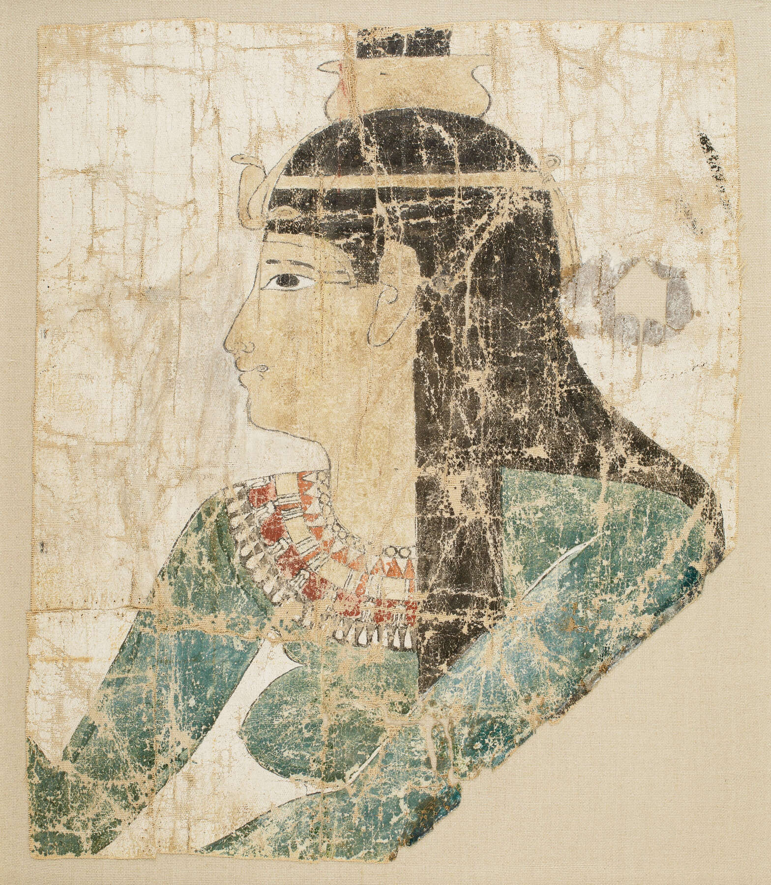 Figure 4.8
Green Pigment Characterization
Egyptian green pigments are notoriously difficult to characterize—and visually identify—for several reasons. First, the aging and darkening of can cause blue pigments to appear increasingly green over time. Another challenge is the fact that certain green pigments (such as malachite, copper chlorides, and organo-copper compounds) can be alteration products, making it difficult to conclude that an area was intended to be green at all. Finally, greens are often the result of pigment mixtures, meaning that multiple components must be characterized in order to make a full identification.
Technical Examination
Because of these challenges, a range of analytical techniques was used to investigate shroud paint surfaces and identify pigments in paint dispersions and cross sections: using Bruker Tracer III-V handheld XRF units with Rd tubes set to 40kV, 12.50μA at 30-second run times with S1PXRF software; using a Hyperion 3000 FTIR spectrometer in transmission mode, with scans from 4000 to 600 inverse centimeters; using a Renishaw system equipped with an He-Ne laser, with an emission line set at 633 nanometers; and finally, using a Philips XL30 environmental scanning electron microscope with Oxford INCA software. The analysis was made possible by the Getty Conservation Institute and the Metropolitan Museum of Art’s Scientific Research Department.
As the research progressed, it became clear how different techniques could be used to answer specific questions about the greens. For example, XRF could narrow the possibilities based on elements present; SEM-EDS and Raman spectroscopy, thanks to their point-identification capability, were especially helpful for identifying the components of mixtures. It should be noted that, subsequent to my research at the Getty, additional analyses were carried out on shrouds 75.AP.87 and 79.AP.219 (see figs. 4.2 and 4.3). They included multispectral imaging, , XRF scanning,13 combined XRF/ analysis,14 and of many areas of color,15 including green.
Multispectral Imaging
was also used to investigate the shrouds’ paint surfaces. Over the past decade MSI has emerged as a valuable tool for examining and characterizing paint surfaces.16 This photographic technique captures the characteristic reflectance, absorption, and luminescence properties of pigments and other materials in an image. Because it can be carried out using a modified DSLR camera, MSI is also a relatively accessible tool in terms of cost.
The imaging method used to examine the shrouds was adapted from a technical imaging manual developed at the British Museum. This manual provides step-by-step instructions for capturing images in visible, and , , and modes, as well as guidelines for image calibration.17 A Nikon D70s UV/IR camera altered to record the electromagnetic spectrum between 250 and 1100 nanometers was used for image capture, as were light sources, lens filters, and calibration standards recommended in the British Museum manual. An open-source calibration workspace developed by the manual’s authors was used to generate standardized multispectral images that can be more readily compared with those obtained under different conditions.
A set of reference paint-outs was created and imaged to provide a centralized visual record of the reflectance, absorption, and luminescence properties of known ancient pigments and binders. The paint-outs have been a useful tool for interpreting the MSI images of artifact paint surfaces and for isolating the particular imaging characteristics of copper-, earth-, and -based green pigments in various binding media. Certain imaging techniques proved especially valuable in the investigation of the shrouds. For example, one could often differentiate between copper-based greens and green earths by the relative darkness of copper greens in UVL images,18 compared with green earths. The presence of a red infrared false color, along with the absence of luminescence in VIL images in green areas on shrouds X.491A and X.491B (see figs. 4.6 and 4.7), indicated that this pigment mixture included indigo. Used in this way, MSI provided important initial information that guided subsequent analysis and data interpretation.
Results
The results of the scientific investigation of the shrouds are summarized in figure 4.9. Information gathered by project collaborators and other scholars is cited in the text.
Shroud | Analysis | Characterization |
|---|---|---|
75.AP.87 | XRF: Ca, Fe, Pb, As peaks XRF mapping: high As counts in leaves19 SEM-EDS: Ca, Si, S, Pb, and As identified in individual pigment particles in green paint cross section Raman: indigo and orpiment identified in point ID of blue and yellow areas of sample cross section20 PLM, infrared imaging confirm presence of indigo, orpiment in leaves21 | Vergaut |
79.AP.219 | XRF: Fe, Pb, As peaks XRF mapping: high As counts in drapery above bird22 FORS: indigo detected23 Infrared imaging indicates presence of indigo in green section of drapery, suggesting that indigo and orpiment were layered to form green24 | Indigo and orpiment, layered or in a mixture |
X.390 | XRF: n/a FTIR: calcite and celadonite25 Imaging: pale blue areas of leaves contain Egyptian blue; green sample area has IRRFC similar to Cyprus green earth (celadonite)26 | Green earth (celadonite?) |
25.184.20 | XRF: Ca, Fe, As, Pb (As may be Pb L alpha peak) FTIR: calcite and celadonite27 Imaging: pale turquoise areas have muted blue IRRFC similar to Cyprus green earth (celadonite); low absorption of same areas in UVL image suggests that pigment is not copper based | Green earth (celadonite?) |
X.491A–B | XRF: Ca, Fe, As FTIR: calcite, indigo, Egyptian green (?);28 orpiment (peak at 800 cm-1)? Imaging: dark green squares are red in IRRFC, consistent with indigo; yellow-green squares painted over background have pale yellow IRRFC, consistent with reference orpiment paint-outs | Vergaut? |
66.99.141 | XRF: Ca, Fe, Cu, As, Pb (As may be Pb L alpha peak) FTIR: Egyptian blue29 VIL imaging confirms that “green” dress is Egyptian blue | Darkened Egyptian blue |
For the Getty objects, XRF analysis of leaves in shroud 75.AP.87 (see fig. 4.2) showed peaks for calcium, iron, lead, and—notably—arsenic, the results of which are confirmed in X-ray maps acquired from the shroud subsequent to this project.30 SEM-EDS analysis identified both sulfur and arsenic in individual pigment particles in a green paint cross section taken from a leaf. An additional paint cross section from the same area revealed particles of and indigo,31 confirming that the green pigment used in the wreath is vergaut, a mixture of the organic blue pigment indigo and the arsenic-sulfide mineral orpiment.32
Shroud 79.AP.219 (see fig. 4.3) was analyzed with XRF in the green-colored middle section of the wing to the boy’s right (fig. 4.10). This analysis yielded XRF peaks for iron, lead, and arsenic. High levels of arsenic were also detected in XRF maps of both the yellow and green sections of the wing.33 FORS analysis conducted subsequent to this research detected indigo in the lower section of the wing, indicating that a combination of indigo and orpiment was likely used to create the green color.34 The green itself appears to be a result of overlap between a stripe of orpiment and a stripe of indigo, although it could also be a mixture of the two pigments; further analysis—such as Raman spectroscopy of a paint cross section—would be needed to confirm.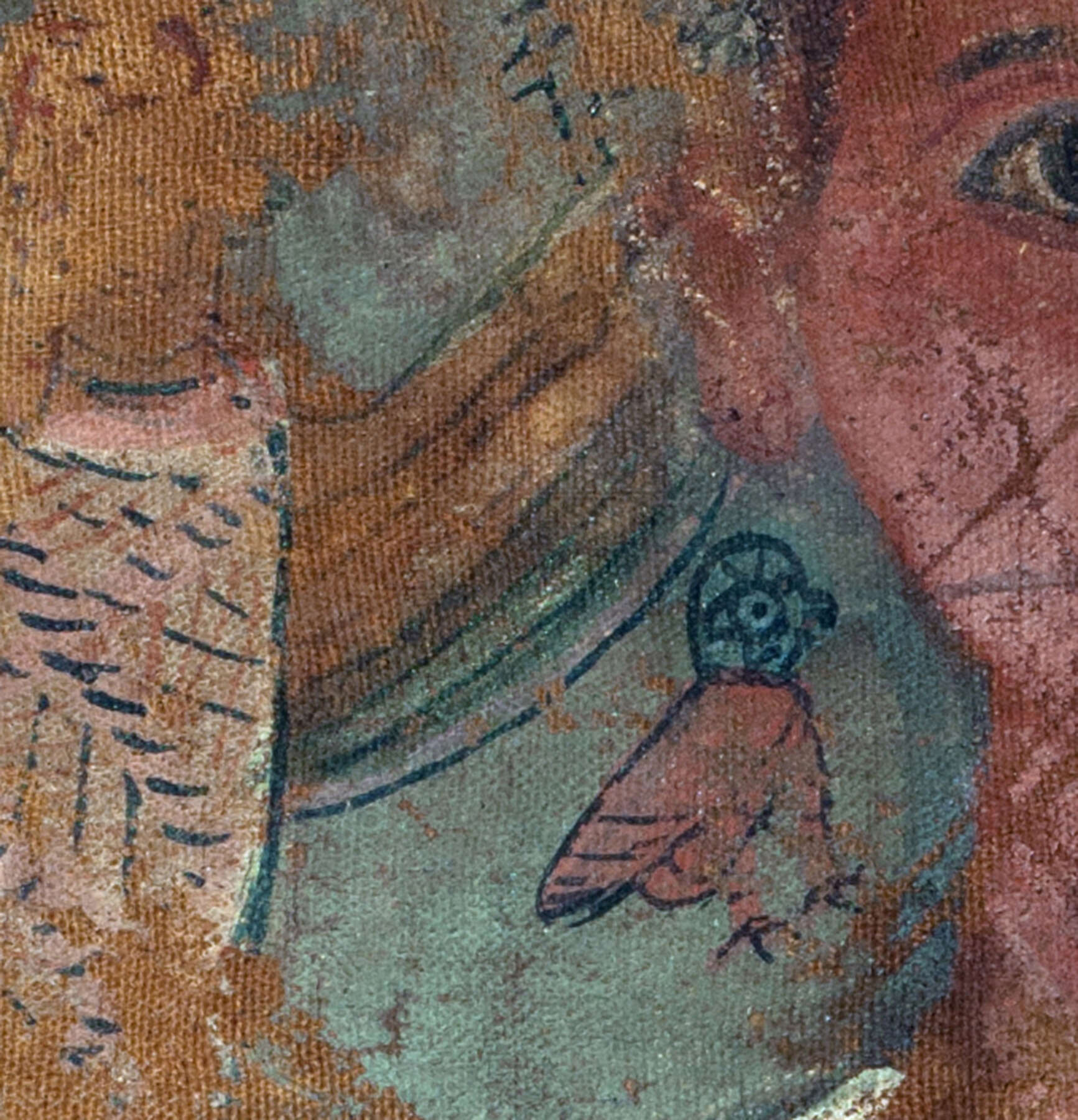 Figure 4.10
At the Metropolitan Museum, a sample of green paint was taken from the garland on shroud X.390 (see fig. 4.4). FTIR analysis yielded a good match for green earth—particularly celadonite.35 This match was further supported by the technical imaging results, in which the infrared false color of the sample area is similar to reference false colors of Cyprus green earth, which has been previously characterized as being predominantly celadonite.36
The Osiris shroud 25.184.20 (see fig. 4.5) was analyzed with XRF in one of several turquoise painted areas. Interestingly, no copper was found in the XRF spectra, which ruled out the possibility that this could be a green frit. FTIR analysis instead suggested that the turquoise color is green earth, a pigment that can carry a wide range of hues from olive green to pale turquoise. The best match to this shroud’s sample was, as in the previous case, celadonite. The imaging supports this result, as does the relatively low absorption of the turquoise areas in the UVL image.
The results for shroud X.491A and B (see figs. 4.6 and 4.7) are less straightforward, but the materials present suggest that the artist used a mixture to produce green. The first clue was the significant arsenic peak in the XRF spectrum taken from a dark green area in the checkerboard pattern behind and to the right of the figure (see fig. 4.11). Multiple FTIR spectra were gathered from a sample taken from the same dark green square. Although one such square indicated a possible match for Egyptian green, the lack of copper in the XRF spectrum acquired in the same area likely rules out its presence. A second FTIR spectrum was generated from the same sample, one that indicated a match for indigo. Could this, in fact, be a mixture of indigo and a yellow pigment? The imaging supports this interpretation, as the green area in question is red (indicating indigo) in the IRRFC images; it also suggests that a layer of orpiment was applied over this dark green mixture to create the light green squares in the same checkerboard pattern. These areas appear bright pale yellow in the IRRFC image—as do other areas of bright yellow color on the portrait. Follow-up analysis with Raman spectroscopy would help to confirm this hypothesis.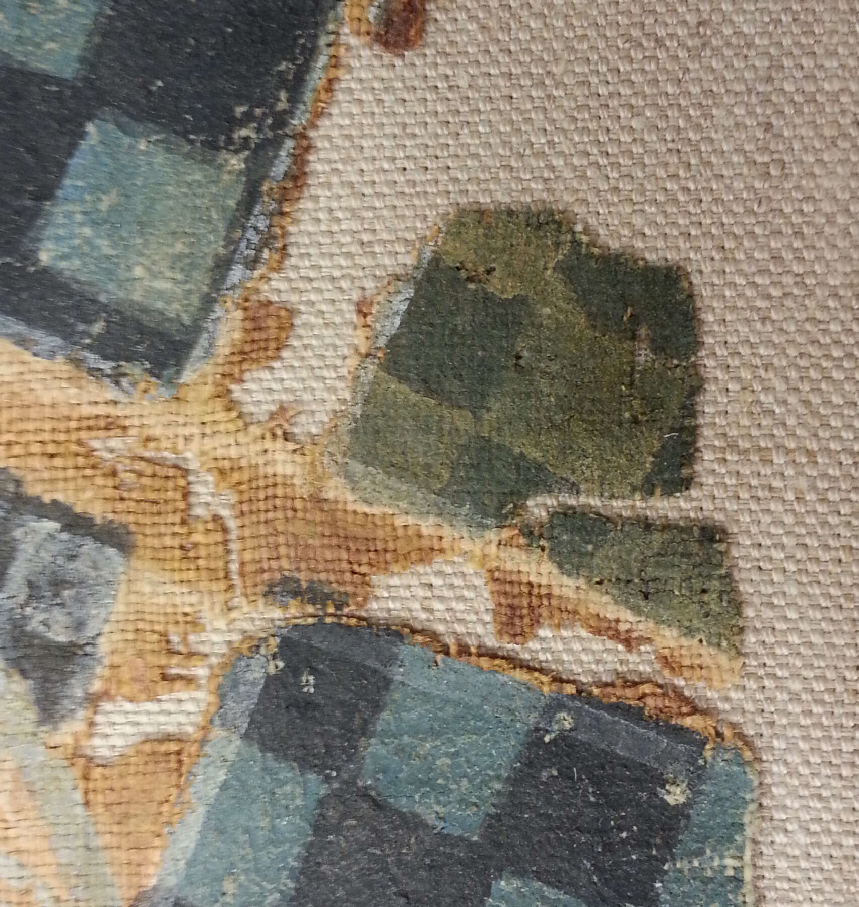 Figure 4.11
The FTIR results from shroud 66.99.141 (see fig. 4.8) strongly indicate that the goddess’s “green” dress is, in fact, a discolored Egyptian blue. VIL imaging showed a strong infrared luminescence throughout the dress, helping to confirm the spectroscopic results.
Discussion
The technical investigation of the shrouds and of other artifacts included in the survey demonstrates how accessible imaging tools can optimize the investigative potential of traditional analytical techniques. In this study, multispectral imaging and analysis informed each other; together, they not only helped confirm ambiguous characterizations but also provided a broader impression of how pigments were used across an artifact’s surface. These combined techniques were especially useful in analyzing the shrouds, where green, blue, and yellow pigments are often mixed and layered.
The conflicting results seen with fragments X.491 A and X.491B (see figs. 4.6 and 4.7) help illustrate the material and chemical complexity of green pigments and mixtures. Although the FTIR spectrum indicates a possible match for Egyptian green in the sampled area shown in figure 4.11, the lack of copper in the XRF spectrum acquired in the same area rules out its presence. Likewise, peaks at 1627, 1462, and 1076 inverse centimeters appear to be from indigo, while a peak near 800 inverse centimeters in spectra from both dark and light green squares could be from orpiment, the latter being consistent with the significant XRF peak for arsenic in the same area. Raman spectroscopy would be needed to confirm that this color is, in fact, a mixture of indigo and orpiment (vergaut).
While questions remain about the exact character of two of the shrouds’ green pigments, it is noteworthy that the only copper-containing pigment found on the shrouds was a discolored Egyptian blue that appeared green. Although copper-based greens continued to be used after the dynastic periods, their absence on the shrouds is interesting and reflects a diversification in green pigment use during Egypt’s Ptolemaic and Roman periods. This shift in pigment selection is suggested not only by the use of vergaut but also by green earth pigments—their best-known mineral deposits were located outside of Egypt,37 and their use on these artifacts offers intriguing physical evidence of extensive pigment trade networks within the Hellenistic and Roman worlds.
Conclusions
The technical study of the painted mummy shrouds provides a small but meaningful addition to a growing Egyptian pigment data set. These and other studies are helping to create a more inclusive knowledge base on Egyptian materials and technology—one that focuses increasingly on the Ptolemaic and Roman periods. The research here demonstrates the sheer variety of greens that may be encountered on an artifact as well as the range of analytical tools that are required for green pigment identification. These analyses also show how pigment choices expanded during Egypt’s Ptolemaic and Roman periods.
Plotting the results of this project’s technical survey graphically (fig. 4.12), helps visualize this shift in green pigment use. In comparing the types of green found on Ptolemaic and Roman Egyptian artifacts with earlier uses of green, one can readily see an expansion in the types of green pigments and combinations of pigments used to achieve the color. Occurrences of green earth become much more common in the Roman period and at this time are found on artifacts more frequently than copper-based greens. Mixtures, likewise, occur more frequently, and mixtures of indigo and yellow occur exclusively during the Roman period.
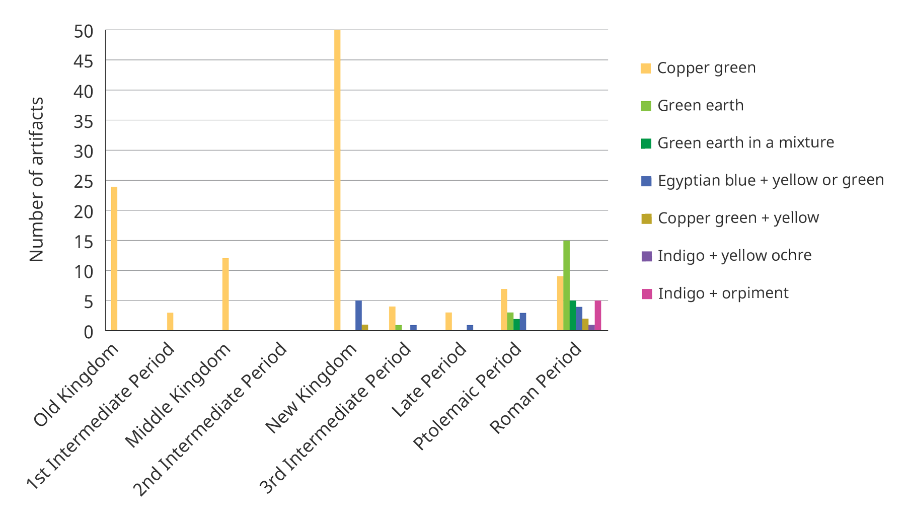 Figure 4.12
This expansion of the green pigment palette coincides with the introduction of traditionally Hellenistic and Roman pigments into Egypt, such as rose and .38 Green earth, too, originates in the Hellenistic paint tradition, and its use appears well established by the time portrait panels and shrouds were in demand.39 The use of blue and yellow mixtures, although present in earlier dynastic Egyptian technical studies, appears to accelerate during the Ptolemaic and Roman periods. With an increasingly diverse palette came many possible green hues and shades and, as seen on a number of the shrouds, a propensity for mixing, layering, and experimenting with color. In portraiture in general, this nuanced use of color is reflected in both artists’ painting techniques and choice of materials.40
The appearance of vergaut is also interesting (fig. 4.13). Perhaps best known for its use in Islamic and Western European illuminated manuscripts, vergaut is mentioned in Eraclius’s tenth-century treatise De coloribus et artibus Romanorum and has appeared on many painted wooden artifacts and shrouds from Egypt and other Roman provinces.41 More examples are sure to emerge as scholars continue to study artifacts from this syncretic period of Egyptian culture; making this evidence widely available is crucial to building complete pigment characterizations, understanding material choices and changes in artistic practice, and providing more, much-needed, technical information about Greco-Roman Egyptian art.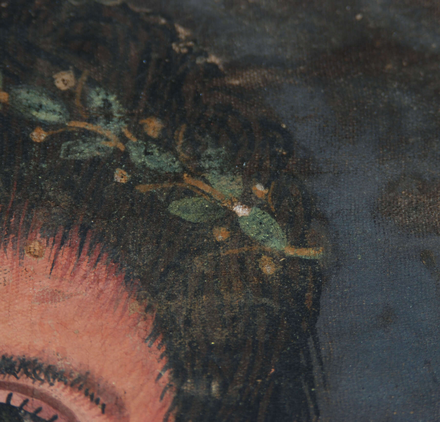 Figure 4.13
Acknowledgments
This research is the product of the collaborative effort of many individuals who have lent their time, expertise, and support to the project over many years. For supervising my fellowship research on green pigments I would like to thank Suzanne Davis and Claudia Chemello at the Kelsey Museum of Archaeology; Marie Svoboda and Jerry Podany at the J. Paul Getty Museum; and Ann Heywood at the Metropolitan Museum of Art. I would also like to thank Emilia Cortes and the Metropolitan Museum’s Egyptian Art Department and Greek and Roman Art Department for supporting my research on the shrouds. I am indebted to many scientists for their assistance and analysis at both the Getty Conservation Institute and the Metropolitan Museum’s Department of Scientific Research, specifically Julie Arslanoglu, Federico Caro, Ilaria Ciancetta, Giacomo Chiari, Art Kaplan, Elisa Maupas, Adriana Rizzo, Karen Trentelman, and Marc Walton. Many thanks as well to Monica Ganio and David Strivay for making the results of their subsequent analyses of the Getty shrouds available. I must also thank Joanne Dyer for her MSI expertise, as well as Anna Serotta and Dawn Kriss for their ongoing collaborative support with reference standards, VIL methodology, and more. Finally, many thanks to J. P. Brown, Laura D’Alessandro, Emily Heye, and Tina March for accommodating my research at their museums.
Notes
- A sample of this pigment was identified as celadonite using a Rigaku Ultima IV X-Ray Diffractometer, at an angle of 2 to 70 degrees, 0.02-degree sampling width, and scan speed of 1, courtesy of the University of Michigan Earth and Environmental Sciences Department.↩
- ; ; ; ; ; .↩
- ; ; ; .↩
- ; ; ; ; ; .↩
- ; ; ; .↩
- ; .↩
- See for full bibliographies of the shrouds; .↩
- .↩
- .↩
- ; .↩
- .↩
- .↩
- .↩
- .↩
- .↩
- ; ; .↩
- .↩
- A phenomenon also observed in .↩
- .↩
- .↩
- ; images courtesy of the J. Paul Getty Museum, UCLA/Getty Program in Archaeological and Ethnographic Conservation, and the Los Angeles County Museum of Art.↩
- .↩
- .↩
- Images courtesy of the J. Paul Getty Museum, UCLA/Getty Program in Archaeological and Ethnographic Conservation, and the Los Angeles County Museum of Art.↩
- .↩
- .↩
- .↩
- .↩
- .↩
- .↩
- .↩
- ; .↩
- .↩
- .↩
- .↩
- .↩
- .↩
- ; ; .↩
- .↩
- .↩
- ; ; .↩