ChicagoVak, Bettina, Roberta Iannaccone, and Katharina Uhlir. “Nondestructive Studies of Ancient Pigments on Romano-Egyptian Funerary Portraits of the Kunsthistorisches Museum, Vienna.” In Mummy Portraits of Roman Egypt: Emerging Research from the APPEAR Project, edited by Marie Svoboda and Caroline R. Cartwright.
Los Angeles: J. Paul Getty Museum, 2020. https://www.getty.edu/publications/mummyportraits/part-two/15/.
MLAVak, Bettina, et al. “Nondestructive Studies of Ancient Pigments on Romano-Egyptian Funerary Portraits of the Kunsthistorisches Museum, Vienna.” Mummy Portraits of Roman Egypt: Emerging Research from the APPEAR Project, edited by
Marie Svoboda and Caroline R. Cartwright,
J. Paul Getty Museum, 2020. https://www.getty.edu/publications/mummyportraits/part-two/15/. Accessed DD Mon. YYYY.
In 2014 the conservation department of the Kunsthistorisches Museum, Vienna (KHM), joined the APPEAR project by adding ten mummy portraits to the collaborative study. Preliminary scientific investigations of these works began at the KHM in 1999.1 Conservation treatment was completed on all ten portraits as well as on five examples from the collection of antiquities of the Museum of Fine Arts, Budapest, to learn more about the acquisition history of the Budapest funerary portraits.2
Conservation scientist Dr. Roberta Iannaccone implemented noninvasive multispectral imaging (MSI)Citation: Multispectral imaging (MSI) / multiband imaging (MBI). The creation of a series of images, each recording reflectance and luminescence within a different limited range of wavelengths. This process involves using a series of band-pass camera filters or a set of narrow-band illumination sources; thus, it records variations in the absorption of materials at different wavelengths. Comparing or combining these images can help to characterize materials or to distinguish between materials that may appear similar. for preliminary pigmentCitation: Pigment. A colorant either derived from natural sources—mineral, plant, or insect—or produced synthetically. Typically, pigments are crushed into a fine powder and mixed with a binder, resulting in a suspension that becomes insoluble when dry; a dye produces a lake pigment when attached to an inorganic substrate or mordant. identification. Dr. Caroline Cartwright, a wood anatomist, identified the tree species used for all fifteen panelsCitation: Panel. Painting support made from various woods, including lime, sycomore fig, and cedar of Lebanon, among others. The shape of the upper portion of mummy portrait panels may indicate the cemetery in which the mummy was buried: stepped panels are associated with Antinoöpolis, round-topped panels with Hawara, and angled panels with er-Rubayat.; for the KHM examples, the results confirmed that six were sycomore fig, three were linden, and one was tamarisk.
Methods
MSI is a set of techniques based on photography at various wavelengths; every range of wavelengths (from ultraviolet to near infrared) can reveal different characteristics.3Ultraviolet-reflected (UVR)Citation: Ultraviolet reflectance or ultraviolet reflected (UVR) imaging / reflected ultraviolet (RUV) imaging. An imaging technique that records variations in reflection and absorption of ultraviolet (UV) radiation by the surface of a subject. This imaging technique primarily aids in the characterization or differentiation of materials. Also because UV radiation exhibits very limited surface penetration, the technique can also help in characterizing surface sheen., ultraviolet-induced visible luminescence (UVL)Citation: Ultraviolet-induced visible fluorescence (UVF) / UV-visible fluorescence / Ultraviolet-induced visible luminescence (UVL) (historically UV/VisFL). An imaging technique and diagnostic examination method, based on characteristic responses of materials to ultraviolet (UV) radiation (185–400 nm) in the form of fluorescence, in which radiant energy in the UV region is absorbed and then reemitted as lower-energy visible light. The fluorescences revealed by the technique are used to assist in the general characterization or differentiation of materials—such as pigments, coatings, binders, and adhesives—and to diagnose the condition of an object (e.g., to detect restorations). The term luminescence also encompasses the possibility of a phosphorescent response to UV radiation in which there is a delay in the reemission of the absorbed energy by some materials, so that emission might even continue for a period after the UV excitation source is turned off. Because fluorescence is by far the dominant phenomenon being observed and documented, the term fluorescence has historically been used in describing this technique in conservation (as well as in medicine, nondestructive testing, and forensics); however, luminescence is an equally appropriate descriptor., ultraviolet-reflected false color (UVRFC)Citation: False-color ultraviolet (FCUV) / ultraviolet-reflected false color (UVRFC). Images created through digital post-processing by combining visible and ultraviolet reflectance (UVR) images. The false colors produced can help in characterizing materials or in distinguishing between visually similar substances., visible (VIS), visible-induced infrared luminescence (VIL)Citation: Visible-induced infrared luminescence / visible-induced luminescence (VIL). An imaging technique in which visible light is used to induce the emission of infrared radiation (primarily in the near-infrared [NIR] region [700–1100 nm]) by certain materials. It has been used to identify historical blue pigments (principally Egyptian blue, Han blue, and Han purple) as well as many cadmium pigments and some natural dyes. These materials may show a very strong IR emission when excited by visible light. The setup for this type of imaging requires an excitation source emitting only visible light with no IR component, an imager with sensitivity to NIR (such as an IR-modified digital camera), and a lens filter that absorbs all visible light and transmits NIR., reflected near-infrared (NIR) photographyCitation: Reflected near-infrared (NIR) photography. An imaging technique that records radiation responses in the near-infrared region (700–1100 nm), thus capturing the contrast between materials that reflect the infrared and those that absorb it, such as carbon-containing pigments. Because infrared is of longer wavelength than visible light, some low-absorbing materials may also allow the infrared to be transmitted through them, revealing hidden underdrawings, artist’s modifications and methodology, or modern interventions., and infrared-reflected false color (IRRFC)Citation: False-color infrared (FCIR) / infrared-reflected false color (IRRFC). Images created through digital post-processing by combining visible and near-infrared images. The false colors produced can help in characterizing materials or in distinguishing between visually similar substances. imaging were used to characterize pigments and shed light on modern retouches.
To acquire the images two cameras were used: a Nikon D80 with a resolution of 10 megapixels and a 23.5-by-15.7-millimeter sensor dimension, and a modified Nikon D3200 with a resolution of 24.2 megapixels with removed IR filter and a 23.5-by-15.7-millimeter sensor dimension. Both cameras were equipped with a Nikkor AF 28–105-millimeter, f/3.5–4.5D lens as well as specific filters for every technique.4
Noninvasive portable micro-X-ray fluorescence analysis (µ-XRF)Citation: X-ray fluorescence (XRF) spectroscopy. A technique used for nondestructive elemental analyses of inorganic materials, utilizing a focused beam of X-rays to excite the atoms on the surface of an artwork and measuring the emitted energy. These emissions provide characteristic fingerprints of the elements in the sampled area, allowing researchers to formulate hypotheses about the compounds contained therein. was performed using the KHM’s PART II instrument.5 The spectrometer, equipped with a vacuum chamber to reduce the absorption of low energetic radiation in air, possesses a low-power X-ray tube with molybdenum (Mo) target (excitation parameters used: 40 kV, 0.4 mA, 100s). The primary beam is focused via a polycapillary lens (spot size ~150 µm). The measuring point is placed 1 millimeter outside of the chamber, thus minimizing absorption losses in the excitation and µ-XRF radiation paths.
Results
For all fifteen investigated portraits the common use of earth pigmentsCitation: Earth pigments. Naturally occurring minerals that contain metal oxides, principally iron and manganese, and that have been used since prehistoric times as pigments. The primary types are ochre, sienna, and umber. was confirmed. Additionally, lead whiteCitation: Lead white. A white pigment, both found as a naturally occurring mineral known as hydrocerussite and produced synthetically by exposing metallic lead to an acid (e.g., vinegar). Lead white has been widely used in antiquity and in Egypt since around 400 BC. Chemical formula: Basic lead (II) carbonate, 2PbCO3·Pb(OH)2, orpimentCitation: Orpiment. An orange-yellow pigment with large particles and a glittering quality used to imitate gold. Sourced from the Red Sea and Asia Minor, orpiment, mentioned by Pliny and Vitruvius and also noted in Egyptian works of the Pharaonic period, was widely traded by the Romans. Chemical formula: Arsenic trisulfide, As2S3 or realgarCitation: Realgar. Closely related to orpiment, a red-orange pigment that was widely traded in the Roman Empire and used throughout ancient Egypt and Mesopotamia. Pararealgar is formed when realgar is exposed to light (degradation); it has the same elemental composition but different crystalline structure. Chemical formula: Arsenic sulfide, As4S4, copper green, Egyptian blueCitation: Egyptian blue (cuprorivaite). A pigment that was manufactured and used by Egyptians possibly as early as 3100 BC. Considered to be the first synthetic pigment, Egyptian blue was made by mixing a calcium and copper compound with silica/quartz and a flux, heating the mixture to a very high temperature (900°C), and then grinding the glassy product to a powder. Chemical formula: Calcium copper silicate, CaCuSi4O10 or CaOCuO(SiO2)4, madderCitation: Madder. A dyestuff derived from the root of the madder plant (Rubia tinctorum), which is native to the eastern Mediterranean and Persia. Likely introduced to Egypt by the Greeks or Romans, madder was used throughout antiquity for coloring textiles and as a pigment. Chemical name: Alizarin (1,2-dihydroxyanthraquinone), Purpurin (1,2,4-trihydroxyanthraquinone), and gold were detected. Also identified were three different types of groundCitation: Ground (preparation layer). A primary layer applied to a substrate to form a smooth surface on which to paint. Typically, ground layers were composed of a white material such as gypsum, although they can range in color and composition. layers: calciteCitation: Calcium carbonate (chalk, lime, calcite). A chemical compound used to create a stable white pigment with limited hiding power (opacity); this pigment is used to make grounds (preparation layers) for painting. Chemical formula: CaCO3, gypsumCitation: Gypsum (calcium sulfate dihydrate). A soft sulfate-based mineral found in nature. Often mixed with water to form plaster, it is used in the preparation of substrates, such as wood panels for painting. Also used as a white pigment, gypsum was identified in Tutankhamen’s paint box. Chemical formula: CaSO4·2H2O, and lead white.
For the purpose of this publication, the studies of two portraits—visually classified as encausticCitation: Encaustic. A wax-based painting technique. From the Greek word enkaustikos (“burned in”), the term in its most literal sense refers to the use of molten beeswax combined with pigments; once solidified, the paint can be further manipulated by the use of heated tools. The term is often used in a more general sense to describe any painting technique in which wax is the major component of the medium. and temperaCitation: Tempera. In the context of ancient art, this term generally refers to a fast-drying, water-miscible painting medium such as animal glue or plant gum. The term tempera originates from the Latin temperare (“combining, blending”). based—are described below.
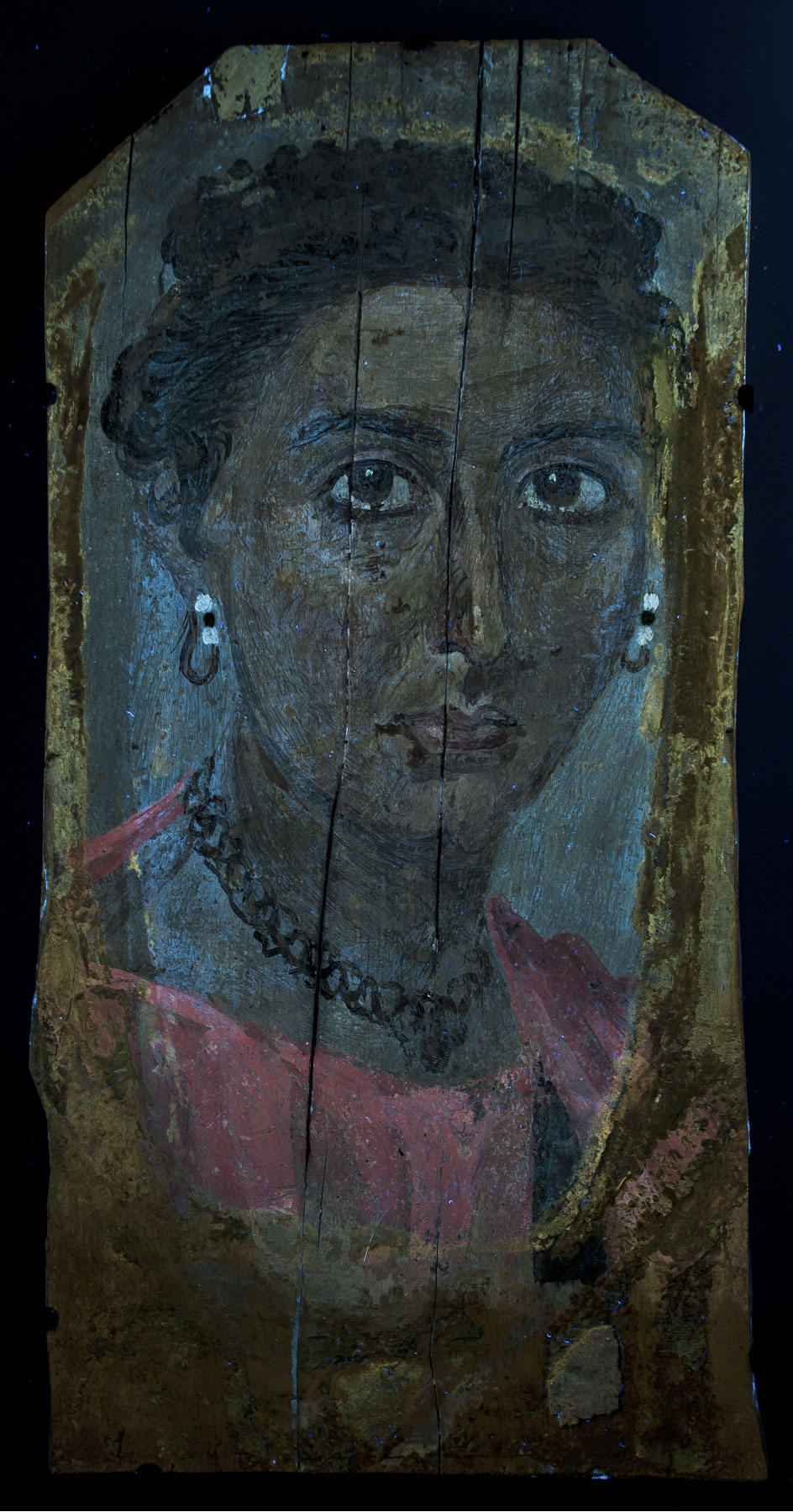
The Portrait of a Lady (fig. 15.1) is encaustic based. The characteristic pink luminescence attributed to a red lakeCitation: Lake. A pigment manufactured by precipitating a dye onto an inorganic substrate/mordant (such as the metallic ions aluminum or calcium). pigment is visible on the tunicCitation: Tunic (chiton). A simple garment that covered the upper body, starting at the shoulders and ending at a length somewhere between the hips and the ankles. The English word chiton originates from the Latin chiton, which means “mollusk”; that, in turn, is derived from the Greek word khitōn, meaning “tunic.” The tunic was a basic garment worn by both men and women in ancient Rome. Citizens and noncitizens alike wore chitons (usually white for men and red for women). Citizens might wear a chiton under the toga, especially on formal occasions. The length of the garment and the presence or lack of stripes (clavi), as well as their width and ornamentation, indicated the wearer’s status in Roman society. but not on the lips; there µ-XRF confirms the presence of iron, suggesting the use of red ochreCitation: Red ochre. A brownish red earth pigment that contains anhydrous iron oxide, or hematite (from the Greek hema, meaning “blood”). Used since prehistory as pigments, ochres may vary widely in shades and transparency. Composition: Anhydrous iron (III) oxide, Fe2O3. The NIR image does not show an underdrawing; however, on the lips the outlining pigment seems mixed with a substance that strongly absorbs infrared radiation, suggesting the presence of charcoal in the admixture.
Figure 15.1 Portrait of a Lady, Romano-Egyptian, AD 117–138. er-Rubayat. Encaustic on wood, 40 x 20 cm (15 ¾ x 7 7/8 in.). Vienna, Kunsthistorisches Museum, Antikensammlung, X 297. KHM-Museumsverband
The violet tunic seems to partially absorb infrared radiation, with IRRFC showing a green/grayish response. Aluminum, typically associated with the lake substrate, together with a strong pink luminescence suggests the presence of red lake. Usually, when a red lake paint layer is superimposed on or mixed with red ochre or cinnabarCitation: Cinnabar. An orange-red pigment with excellent hiding power (opacity) and good permanence. It has been used from antiquity to the present. Chemical formula: Mercuric sulfide, HgS, it appears yellow/orange in IRRFC. The VIL image shows the typical luminescence of Egyptian blue in areas of the tunic. µ-XRF measurements confirm this observation, revealing some copper. Most likely, the red lake was combined with Egyptian blue, lead white, and red ochre (detected by µ-XRF) to obtain the purple tint. Egyptian blue was mainly used to render the face and create shadows (fig.15.1c).
Identification of the dark blue clavusCitation: Clavus (pl. clavi). A vertical stripe or ribbonlike ornament, placed in pairs, that adorned the shoulders of a tunic. In Rome some clavi of specific width and/or color distinguished members of particular rank or status, but the significance of the clavus in an Egyptian context remains undetermined. on the left side of the tunic was inconclusive. XRF analysis did not identify any characteristic elements, and UVRFC and IRRFC imaging techniques did not indicate the presence of indigoCitation: Indigo. A natural blue dye derived from the plant Indigofera tinctoria and related species growing in the Mediterranean, India, and Asia, among other locations. It is believed that originally the dye woad (Isatistinctoria), rather than indigo, was used in antiquity. Chemical formula: C16H10N2O2. Furthermore, a pale blue fluorescence can be observed on top of the painted surface, in areas not covered by the mummy wrappings.6 Classification and origin of this material will be a matter of further investigation.
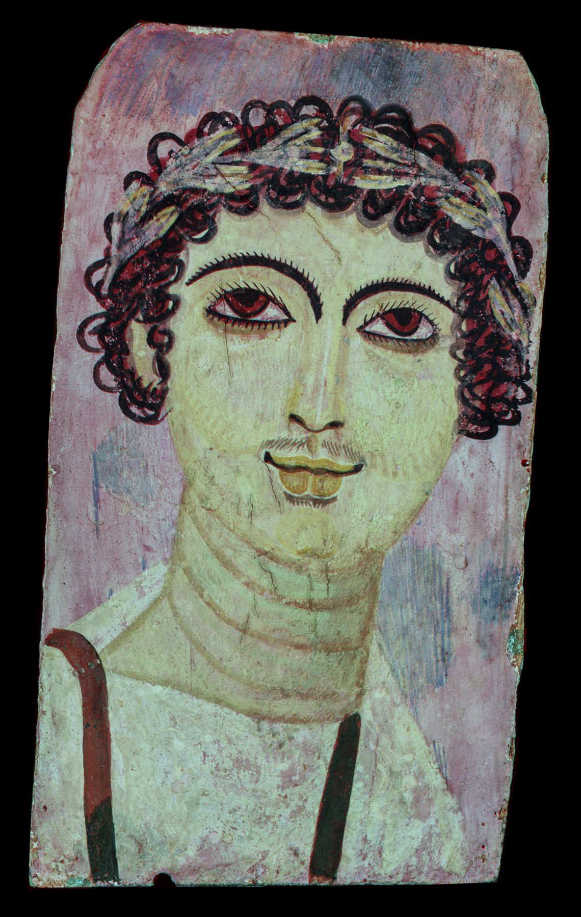
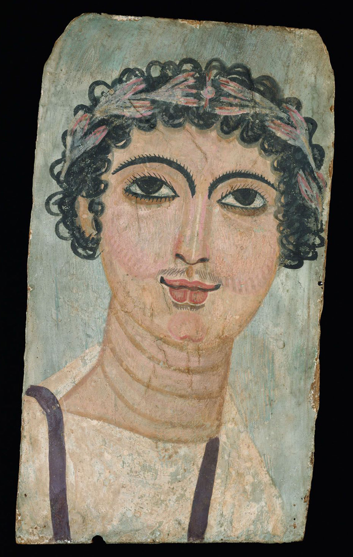
The Portrait of a Young Man with Wreath (fig. 15.2) is tempera based. The UVL image shows some areas with bright pink fluorescence (likely due to a red lake) mainly on the wreathCitation: Wreath. An assortment of flowers, leaves, fruits, twigs, or other materials constructed to resemble a loop. Typically worn on the head in ceremonial events, wreaths have much history and symbolism associated with them. In the Greco-Roman world, wreaths were used as adornments that could represent a person’s occupation, rank, achievements, or status., cheeks, and lips; a bright yellow luminescence can be observed on the bridge of the nose, the lip outlines, and the eye. µ-XRF analysis in the area of yellow luminescence identified the presence of lead (lead white) and iron (earth pigments). Lead white typically fluoresces light blue; therefore, this particular yellow fluorescence could be related to the binder or a pigment mixture.7
Figure 15.2 Portrait of a Young Man with Wreath, Romano-Egyptian, AD 125–150. er-Rubayat. Tempera on wood, 32.5 x 18 cm (12 13/16 x 7 1/16 in.). Vienna, Kunsthistorisches Museum, Antikensammlung, X 432. KHM-Museumsverband
The µ-XRF spectrum of the irises indicates the presence of iron (earth pigment). The unexpected red color visible in the IRRFC image of the dark hair, the original portion of the clavi, the irises, and the light blue background is attributable to the spectral response of a blue pigment, probably an organic blue (indigo) partly mixed with an earth pigment.8
Our understanding of the materials and painting process of the mummy portraits described above has been much enhanced by the scientific identification of the woods and by the use of two nondestructive methods (µ-XRF and MSI) for identifying the pigments.
Notes
Pitthard et al. 2007Citation: Pitthard, Václav, Bettina Vak, Martina Griesser, Sabine Stanek, and Manuela Laubenberger. 2007. “Fayum Portraits from the Collection of Greek and Roman Antiquities, Kunsthistorisches Museum, Vienna: Conservation Treatment and Research into Composition.” Technologische Studien Kunsthistorisches Museum 4: 11–29..
↩
For similar methods applied on Attic ceramics, see Vak 2013Citation: Vak, B. 2013. “Auf der Suche nach dem Original: Anwendungen und Illustrationen naturwissenschaftlicher Diagnostik an attisch figürlich bemalter Keramik aus der Antikensammlung des Kunsthistorischen Museums.” Corpus Vasorum Antiquorum Österreich 1: 41–72..
↩
Buzanich et al. 2010Citation: Buzanich, Günter, P. Wobrauschek, Christina Streli, A. Markowicz, D. Wegrzynek, E. Chinea-Cano, Martina Griesser, and Katharina Uhlir. 2010. “PART II (Portable ART Analyzer)—Development of a XRF Spectrometer Adapted for the Study of Artworks in the Kunsthistorisches Museum, Vienna.” X-Ray Spectrometry 39 (2): 98–102.; Uhlir et al. 2012Citation: Uhlir, Katharina, B. Frühmann, G. Buzanich, M. Griesser, C. Streli, P. Wobrauschek, B. Grossmayer, and S. Smolek. 2012. “A Newly Developed, Portable, Vacuum-Chamber Equipped XRF-Instrument, Designed for the Sophisticated Needs of the Kunsthistorisches Museum, Vienna.” IOP Conference Series: Materials Science and Engineering 37: 1–7. http://iopscience.iop.org/1757-899X/37/1/012008/pdf/1757-899X_37_1_012008.pdf.
↩
For further information about the material, see Pitthard et al. 2007Citation: Pitthard, Václav, Bettina Vak, Martina Griesser, Sabine Stanek, and Manuela Laubenberger. 2007. “Fayum Portraits from the Collection of Greek and Roman Antiquities, Kunsthistorisches Museum, Vienna: Conservation Treatment and Research into Composition.” Technologische Studien Kunsthistorisches Museum 4: 11–29..
↩
See La Rie 1982Citation: La Rie, E. René de. 1982. “Fluorescence of Paint and Varnish Layers (Part I).” Studies in Conservation 27 (1): 1–7.; Roy 1993Citation: Roy, Ashok, ed. 1993. Artists’ Pigments: A Handbook of Their History and Characteristics. Vol. 2. Oxford: Oxford University Press., 67–81.
↩
Indigo has been identified in the pupil, hair, clavi, and beads of a comparable portrait in the Getty collection: Mummy Portrait of a Woman (79.AP.129). The paintings are similar in both their execution and their response to MSI.
↩
 a. UVL
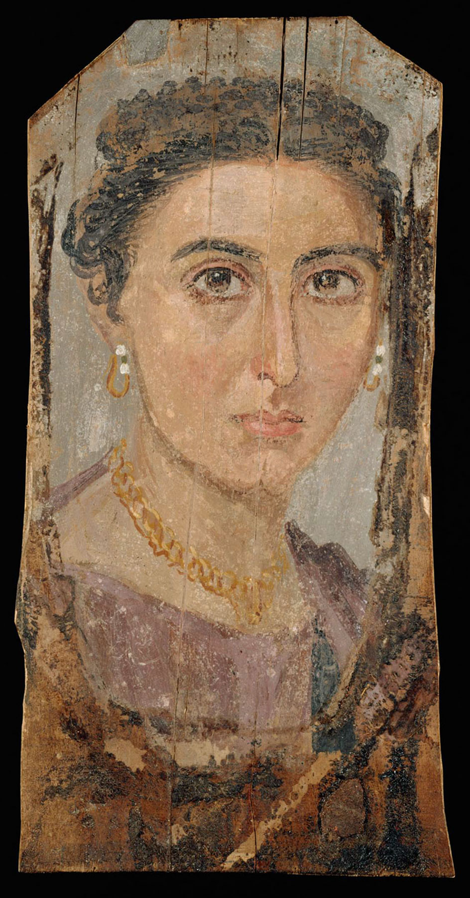 b. VIS
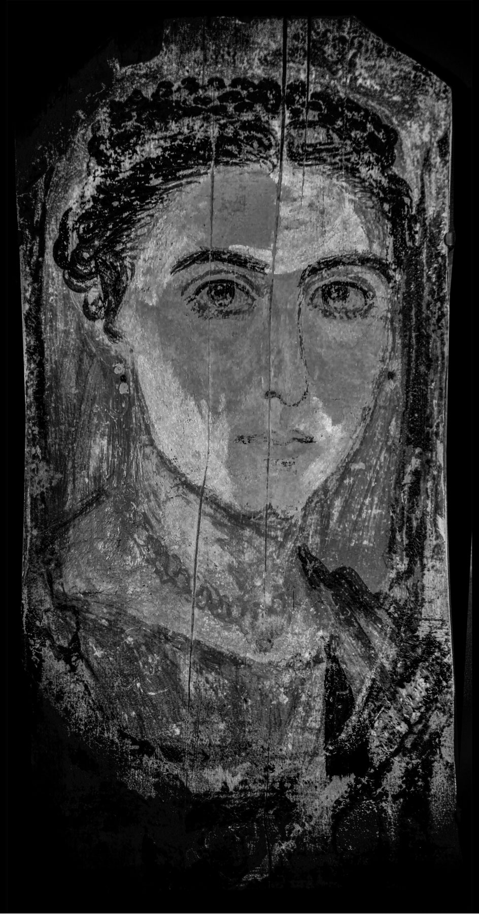
 a. VIS
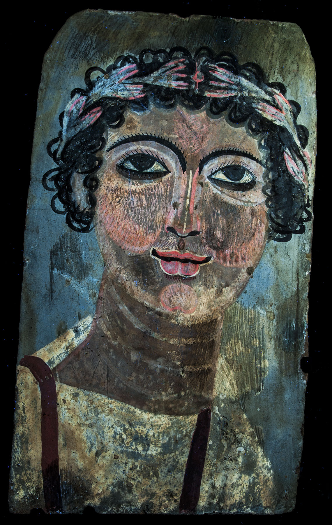 b. UVL
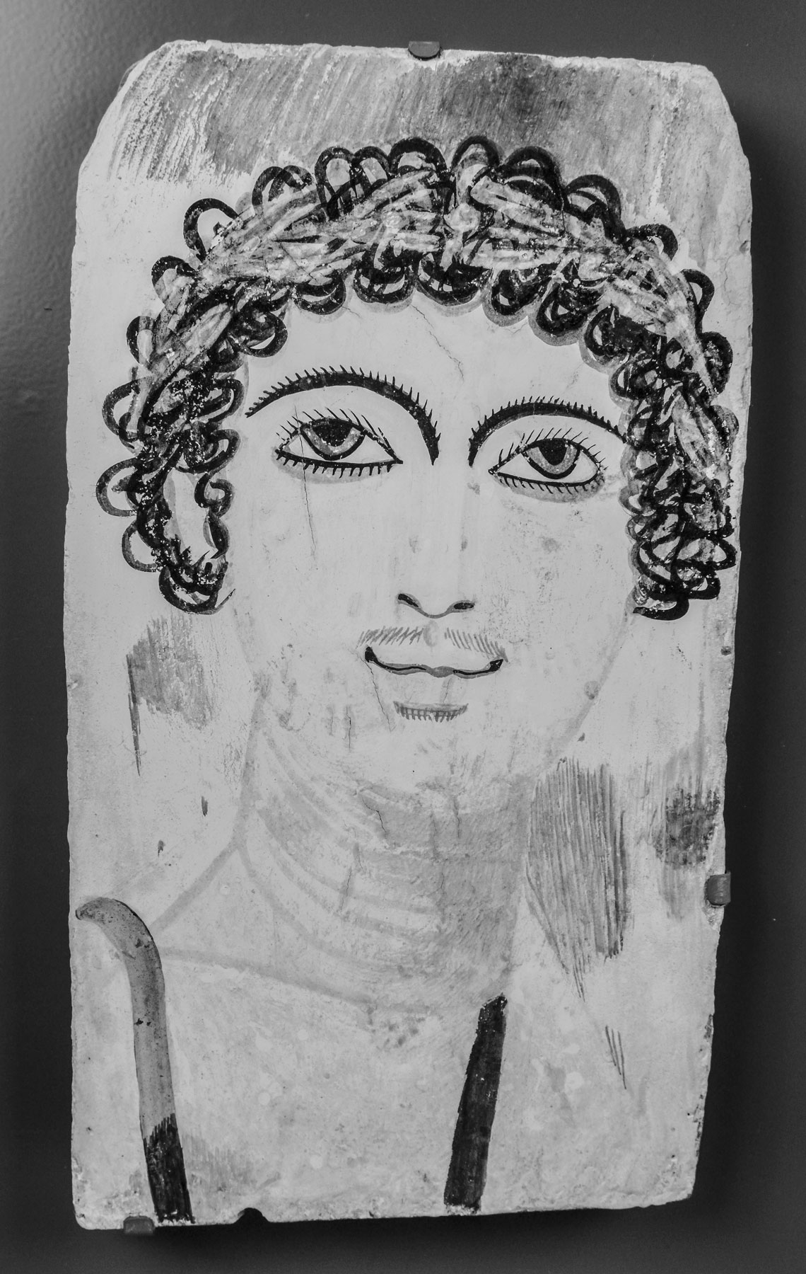 c. NIR